
Following the COVID-19 crisis, a surge in cases of acute kidney failure prompted the Pathology Department to initiate investigations at the Core Facility Cellular Imaging Electron Microscopy Centre Amsterdam. The focus was to identify virus particles and viral proteins in kidney of affected patients, aiming to enhance the understanding of SARS-CoV-2's impact on renal function. This research aimed to refine treatment strategies for hospitalized patients and those experiencing post-COVID kidney complications. The findings, recently published in Microbiology Spectrum, revealed unexpected insights.
Research technician Anita Grootemaat and colleagues, demonstrated that the SARS-CoV-2 virus behaves different than expected. Since the SARS-CoV-2 virus needs all of its proteins to replicate, the researchers were expecting to find all of them in the kidney. The research project included several patients, yet the researchers identified only one instance of the SARS-CoV-2 protein in the kidney of a patient who had recovered from the infection. A single but very stable protein: the nucleocapsid protein has been detected in the epithelium. So either this protein is not degraded and has been accumulating in the kidney, or this protein is still being produced without the other viral proteins. Either way, the immune system of the patient responds to the presence of this strange protein and therefore causes the acute kidney problems the patient faces after recovery of SARS-CoV-2.
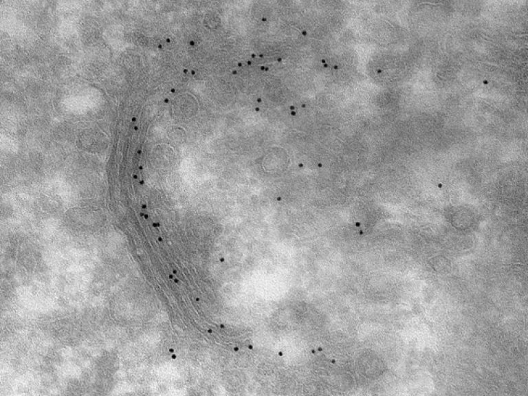
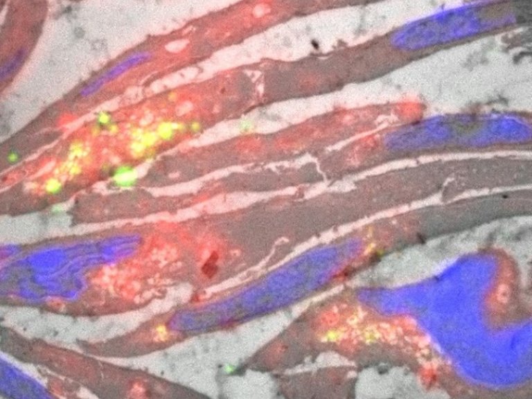
Images: In Left, nucleocapsid protein labelling decorated with gold (black dots) in kidney post-COVID patient. Scale bar 200 nm. Right image of SARS-CoV-2 infected cells imaged with a combination of Fluorescence and Electron Microscopy. In green double strained RNA, in red lipid accumulation, in bleu nucleus and grey the Electron Microscope image of the infected cells. The yellow spots indicate the accumulations of both dsRNA and lipid. Scale bar is 5 μm.
Future perspective
Nicole van der Wel, Head Electron Microscopy Center Amsterdam at Amsterdam UMC: “Towards the future we want to investigate if more patients suffer from Nucleocapsid accumulations in the kidney and if so, how we can avoid or block this accumulation. Furthermore, we want to investigate if the virus replicates in the kidney regions. We have shown in cells infected with SARS-CoV-2 that dsRNA, which is another sign of replication, is present in lipid filled compartments. Also we want to understand the effect of the lipid accumulation as we often detected this close to the Nucleocapsid clusters in kidney but, in previous research link, also in lung of COVID patients”.
For more information, contact Nicole van der Wel or read the scientific publication.
All Figures with interactive mode are available here.
Researchers involved at Amsterdam UMC
Anita E. Grootemaat, Research technician at Amsterdam UMC 1
Niek Wiersma, BSc student Biomedical Sciences at Universiteit van Amsterdam 1
Sanne van der Niet, Post-doc Medical Biology at Amsterdam UMC, 1,3
Irene M. Schimmel, Operator Electron Microscopy at Amsterdam UMC 1
Sandrine Florquin, Professor of Pathology at Amsterdam UMC 2,3
Eric A. Reits, Professor Cellular Imagingat Amsterdam UMC 1
Nicole N. van der Wel, Head Electron Microscopy Center Amsterdam at Amsterdam UMC 1,3
Contact information:
1Electron Microscopy Centre Amsterdam, Medical Biology, Amsterdam University Medical Centre AMC, the Netherlands
2Department of Pathology, Amsterdam University Medical Centers (location University of Amsterdam), Amsterdam, The Netherlands
3Amsterdam Institute for Infection and Immunity, Amsterdam, The Netherlands
Funding
Sanne van der Niet's research is supported by a grant from the National Institutes of Health (NIH).
Explore the previous achievements of Nicole van der Wel, Anita Grootemaat and Sanne van der Niet in our archived news articles:
Text: Esmée Vesseur