PET-CT scanners use gamma-rays from infused radioactive tracers to create a 3D map of the radioactive tracer distribution, thereby visualizing biological information across the body, in organs and/or tumors. With precise and detailed pictures of organs and tissues, PET-CT imaging technology has revolutionized medical diagnosis and advanced precision medicine over the last 20 years.
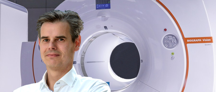
The new Biograph Vision Quadra from Siemens Healthineers is a groundbreaking PET-CT system that is a quantum jump forward in imaging technology. The system is part of the Amsterdam Oncology and Neuroscience Research (ADORE) project.
What does it do better than standard PET-CT scanners?
Larger field of view
The radioactive tracer used in PET scans is administered to a patient and spreads throughout the entire body. However, current imaging system’s detection axial field of views are only up to about 25 cm, meaning physicians can see only a small portion of the body at a time.
The Quadra system is four times larger with a 106 cm axial field of view. This covers ~85% of the body, allowing a view from the top of the head to mid-thigh on an average adult. This 'whole-body' range encompasses all the critical organs mostly evaluated in molecular imaging studies.
“The system enables us to image all major organs at once, immediately after administration of the radiotracer. This is the most important breakthrough,” explains Ronald Boellaard, Professor, Radiology and Nuclear Medicine. “Never before has it been possible to follow radiotracer uptake dynamically over all critical organs in the body. Because we can do this now, we get a lot more information and we can do much more extensive analyses."
Ultrahigh sensitivity & lower radiation exposure
Covering more anatomy in one scan position also directly increases sensitivity by capturing more of the radioactive signal. The sensitivity of the Quadra is about 10 to 20 times higher compared to current state-of-the-art PET/CT systems.
The higher sensitivity and wider view mean that much less radiation is needed. “Using less radiation opens the door to doing more frequent and/or multi-radiotracer repeated PET-CT scans to allow follow-up investigations or a more comprehensive biological characterization of organs, such as the brain, or tumors,” says Prof. Boellaard.
Huge potential for patients and research
Advantages offered by the new system include, but are not limited to:
- Earlier and better detection and diagnosis of cancer or neurodegenerative diseases,
- Investigation of cell-based therapies including trafficking patterns,
- A more comprehensive (whole body) investigation or characterization of oncological and neurological diseases, among others,
- Acceleration of new drug development,
- Rapid clinical deployment of new therapeutic agents.
“It will enable researchers at Amsterdam UMC to be amongst the world’s leaders in molecular imaging and ensures our patients are receiving world-class management of their conditions.”
Q&A with Prof. Ronald Boellaard
Is the scanner operational?
“The system has been in use since mid-May. So far mainly for diagnostic examinations, but the first research studies have started on this system, including my own research into new scanning procedures.”
What are your initial impressions?
“It is now clear that we can obtain much better images with this system. The images are less noisy and sharper, which means we can detect small abnormalities (tumors) in the body or brain much earlier.”
What type of investigations are you personally looking forward to performing?
“The main breakthrough will be dynamic multi-tracer whole body imaging in order to capture more information from a single scan or single examination session, which requires 2 or more separate examinations on current standard systems.
“Another very interesting opportunity would be to measure trafficking or radio-labelled cells, for example CAR T-cells, to obtain better understanding of these novel cell-based therapies.”
Enabling progress
The Amsterdam UMC Imaging Centre and the total body PET-CT system are made possible because of contributions from the European Regional Development Fund (ERDF), the City of Amsterdam, the Department of Economic Affairs of the Netherlands, and the Province of Noord-Holland. It is actively supported by Amsterdam Economic Board and Amsterdam Marketing, Kansen voor West. In addition, the total body PET-CT system, part of the Innovatiecentrum ADORE project, is financially supported by the Amsterdam Oncology and Neuroscience Research (ADORE) institute’.
For more information contact Prof. Ronald Boellaard.
Text by Laura Roy.
This article was created for Cancer Center Amsterdam.
©2022 - NHBC – Your partner in science communication.