Gliomas are one of the most common types of primary brain tumors. Gliomas cannot be fully cured and require regular follow-up care to monitor the effects of treatment and reassess treatment options.
Recently developed molecular genetic diagnostics can refine glioma classification and tailor treatment, but these techniques are still too expensive for routine diagnostics.
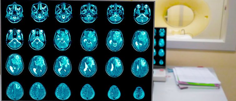
MRI is not using its full clinical potential
Magnetic resonance imaging (MRI) is the current standard for glioma assessment. Current glioma MRI protocols often follow rigid follow-up interval guidelines and involve injection of a gadolinium-based contrast agent to produce contrast enhanced (post-contrast) images. But there are a number of problems with this current procedure:
- The gadolinium-based contrast agent yields inconsistent imaging findings or biomarkers, e.g., falsely suggesting progression or showing progression too late
- Contrast enhancement does not equate to higher glioma malignancy (non-enhancing gliomas can be aggressive)
- The gadolinium-based contrast agent is expensive and potentially harmful to the
patient - The amount of gadolinium-based contrast agent in drinking water is increasing
heavily - Rigid follow-up intervals hinder the accurate detection of volume progression,
leading to under- and delayed reporting of progression - Routine MRI protocols for glioma imaging have not integrated recent advances in MRI techniques
“MRI is not being used at its full potential. In addition, even though there has been much progress in enlisting the power of AI in medical imaging, we are not using that yet in glioma assessment,” says project leader Dr. Vera Keil. “We want to show that a combination of AI, advanced MRI sequences, and a different visual analysis approach can lead to higher diagnostic precision and will in the future make gadolinium contrast injection superfluous for many glioma cases.”
The vision of GLIOCARE
The research project is called GLIOCARE, an acronym for ‘Glioma Imaging Omitting Contrast through Artificial intelligence and Risk Evaluation’. The aims include:
- Scanning 200 prospective glioma patients with advanced MRI techniques (arterial spin labelling and amide-proton-transfer chemical-exchange-saturation-transfer imaging (APT-CEST) protein imaging)
- Developing AI-based methods to assess gliomas based on gadolinium-free images as well as generating synthetic post-contrast images
- Assembling and analysing with AI 1,400 patients’ data in a strong longitudinal glioma MRI database ‘IMAGO’
- Three research fields joining strengths to solve one large clinical problem: Radiology, Neurosurgery, Pathology
- International cooperation with ongoing Cancer Center Amsterdam and Dutch Cancer Society-funded research projects
About Hanarth Fonds
The Hanarth Fonds was founded on September 28, 2018, thanks to the legacy of Arthur del Prado, founder and former CEO of ASM International. The fund aims to promote and enhance the use of artificial intelligence and machine learning to improve the diagnosis, treatment and outcome of patients with cancer.
For more information, contact Vera Keil.
Researchers involved at Cancer Center Amsterdam
Prof. Frederik Barkhof
Dr. Vera C. Keil
Dr. Henk Mutsaerts
Ivar Wamelink (Ph.D. student)
Prof. Pieter Wesseling
Prof. Philip De Witt Hamer
This article was created for Cancer Center Amsterdam.
© 2023 NHBC– All rights reserved.