How was bone marrow diagnostics performed before?
Van den Ancker explains: “We have a pool of analysts who are excellent at assessing which cell is which, and whether they see malignancies. Technician 1 counts and assesses the single cells on the bone marrow smear, and then technician 2 does the same. After the hematologist reviews the bone marrow, the counts and other findings are authorized. This goes fine, but it takes time. Especially with the manual entry of all the data into the laboratory information system (LIS).”
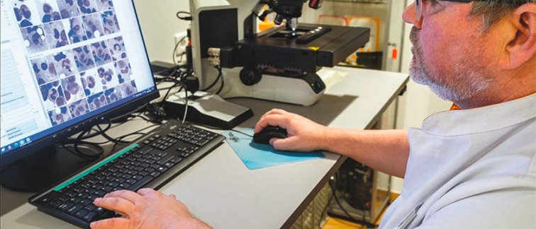
Van den Ancker is therefore pleased with the arrival of a digital microscope for bone marrow diagnostics. “This scans the entire bone marrow smear and then the individual cells of patients suspected of having a hematological malignancy. The microscope then sends the scanned images digitally to the computer. This removes a lot of administrative burden for technicians and clinical chemists. We can therefore focus more on the substantive assessment of the cells and the cohesion of the cells in the bone marrow. That's the fun part of the job.”
Of course, less administrative burden is not the only reason for switching to the implementation of digital microscopy, although it does help reduce the workload for lab analysts amid acute staff shortages.
Diagnostics is also faster now because it is (semi-)automatic. The images from the microscope are digitally processed by the computer resulting in a faster diagnosis. This improves the quality of care, because less time is lost in the diagnostic process.
Multidisciplinary consultation
Faster, more fun, and also more reproducible, because the digital images from the microscope, magnified a thousand times, can be be reviewed and reassessed at any time. Van den Ancker: “This especially helps with bone marrow smears that are difficult to assess. Sometimes analysts have different opinions about which cell is which. Do we all see the same thing, or do we think we see something else? Then it's nice if you can review the cell images together. With manual counts that is a lot more difficult.”
Agreed, adds pathologist Sybren Meijer, who is closely involved in the implementation of digital pathology at the Amsterdam UMC. “The pathologists do the tissue biopsies, the clinical chemists analyze the single cells. The assessment of diagnostic material always somewhat subjective. On Friday afternoon you might count the cells slightly differently than on Monday morning. That's not a bad thing, we are professionals, and intersubjective review always takes place. But it helps if you have the images ready and can look at it together later. You can never achieve one hundred percent objectivity, but you can get close.”
Certainly for the multidisciplinary consultation (MDO) of hematologists, pathologists and clinical chemists it is an advantage that digitized images from the microscope can be shared on the spot, for example on a screen linked to the computer.
Van den Ancker: “Especially in smears of patients about which there are differences of interpretation, that is of added value. The hematologists are therefore enthusiastic about the possibilities of digital microscopy.”
The same applies to sharing digitized images during the training of fellows or physicians in training. Meijer: “This is an improvement in the quality of medical training. In the training rooms, there could be two hundred microscopes and two hundred students taking turns to look at the specimens. That's not only time-consuming, it also means you look at the same specimens two hundred times. Studying digital images together is faster, and you know for sure that you are all looking at the same thing. You thereby increase the learning efficiency.”
Micrometering
Digital microscopy is not new. For the diagnosis and differentiation of blood cells in peripheral blood it has been used in Netherlands for about ten years. That the technique is only now becoming available for bone marrow diagnostics is because you need a much higher resolution to properly assess cells, Van den Ancker explains. “Cells are round, although they are more or less flattened on the smear. That means the microscope has to be able to look through several cell layers. You're not looking at a flat surface, but in a three-dimensional environment where you have to get every cell particle in focus. You do that by continuously zooming in and out with the lens. That's called micrometering. For a long time, the software was not sophisticated enough to make these 3D images, but now it is.”
The microscope has only been in use for clinical care at the VUmc since last October, so experience with it is still accruing. But so far the digital technology is meeting all expectations: speed, less subjectivity, and better reproducibility. Although preparations were necessary to make its arrival possible, Van den Ancker emphasizes. “The link to the LIS took some doing. It is good to take this into account if you want to purchase a digital microscope. Training your staff is also important, although our technicians mastered the technology pretty quickly. And digital microscopy is not cheap, so you have to carefully weigh costs and benefits. But we are satisfied. And interest from other hospitals is strong. Many clinical chemists have already visited us to see the microscope, to see the images, to learn from our experiences. We are the first, but I expect more hospitals will follow.”
Willemijn van den Ancker obtained a PhD for her research at the department of Hematology of the VUmc and subsequently completed her Medicine study. Next, she did a training in clinical chemistry at the OLVG while completing a MBA Healthcare at the University of Amsterdam (UvA). Since 2018, Van den Ancker has been working at the Hematology Laboratory for Bone Marrow and Blood Research at Amsterdam UMC.
Sybren Meijer. After completing his doctoral studies in Portland, Oregon USA, Sybren Meijer finished his training as a pathologist at the Pathology Department of the AMC. He has been working as a pathologist at Amsterdam UMC, and is closely involved in the realization of a completely digitized pathology diagnostic workflow. His research area concerns improved detection and progression prediction of gastroenterology tumors and precursor abnormalities thereof with applied Artificial Intelligence (AI).
This article by Michel van Dijk was originally published in Dutch in Laboratoriumgeneeskunde.
Translated and copied with permission.