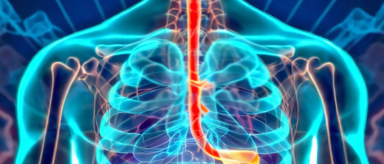
of the esophagus is replaced by tissue similar to the intestinal lining, has long been a challenge in medical endoscopy. Early neoplastic lesions often have a subtle appearance, making them difficult to detect through traditional endoscopic methods. However, a recent breakthrough was achieved by incorporating the assistance of AI for the early detection of neoplasia during endoscopy. The significant advancement is a result of efforts by the Barrett's Oesophagus Imaging for Artificial Intelligence (BONS-AI) consortium, co-led by researchers from Cancer Center Amsterdam, and reported in The Lancet Digital Health.
Barrett's esophagus is a condition that occurs when the normal lining of the esophagus (the ‘tube’ that connects the mouth and stomach) is replaced by a different type of tissue similar to the lining of the intestine. This change is often a response to prolonged acid reflux, where stomach acid irritates and damages the lining of the esophagus.
People with Barrett's have a high risk of developing esophageal cancer. The altered cells in the esophagus can begin to grow abnormally (neoplasia) which can potentially lead to esophageal adenocarcinoma (a type of esophageal cancer). Due to the risk for cancer development, individuals diagnosed with this medical condition, especially those with neoplasia, are closely monitored through regular endoscopies, where a doctor examines the esophagus with a camera.
However, early neoplastic lesions in Barrett's esophagus can be extremely subtle, which makes it difficult for even experienced endoscopists to detect. This increases the risk of missed lesions or delayed diagnosis, increasing the risk of progression to esophageal cancer which, at later stages, has a poor outcome.
The Largest AI Study in the Field
The Barrett's Oesophagus Imaging for Artificial Intelligence (BONS-AI) consortium now reports the successful incorporation of AI into image analysis during endoscopies. The consortium is led by the Department of Gastroenterology and Hepatology, Amsterdam UMC, and the Department of Electrical Engineering, Eindhoven University.
The Computer-aided detection (CADe) system developed by BONS-AI was trained on a diverse dataset from 15 international centers, involving over 14,000 images. The dataset was also meticulously evaluated by 14 Barrett's esophagus experts, ensuring a high level of accuracy and reliability.
“This is the largest AI study in the field so far,” says Jacques Bergman, Professor at Cancer Center Amsterdam of Imaging and Biomarkers, and Director of Endoscopy at the Department of Gastroenterology and Hepatology. “The CADe system was developed using a large, heterogeneous dataset from 15 international endoscopy centers reflecting real-world variability, making it extremely robust.”
Revolutionizing Detection
With CADe assistance, the sensitivity for neoplasia detection among endoscopists increased significantly, from 74% to 88% for images and 67% to 79% for videos. Notably, this improvement did not compromise specificity, which remained high. In the total test set, the CADe detected neoplastic lesions in an impressive 95% of images and 97% of videos. In the benchmarking test set, the CADe system outperformed general endoscopists (90% vs 74%) and proved to be on par with Barrett's esophagus experts (90% vs 87%).
“Our robust CADe is of clear value to the general endoscopists,” says Prof. Bergman. “Moreover, the system is specifically designed for direct implementation into current existing endoscopy platforms. These elements are essential for successful integration into routine clinical practice.”
Reshaping the Medical Imaging Landscape
The study's findings are a testament to the transformative impact of AI in reshaping the medical imaging landscape. The system's superior performance in identifying neoplasia, particularly in its subtle forms, demonstrates its value as a powerful assistive tool for endoscopists. BONS-AI is now working to enhance the graphical user interface for better interpretation by endoscopists and to refine the system to reduce false positives without sacrificing sensitivity. The aim is to implement CADe into routine clinical practice to achieve more accurate diagnoses, timely interventions, and ultimately better patient outcomes.
For more information, watch this video, or contact Prof. Jacques Bergman, or read the scientific publication:
Fockens, K.N., Jong, M.D., et al. A deep learning system for detection of early Barrett's neoplasia: a model development and validation study. The Lancet Digital Health, Volume 5, Issue 12, e905 - e916. DOI: https://doi.org/10.1016/S2589-7500(23)00199-1.
Researchers at Amsterdam UMC
M.R. Jong, co-first author
K.N. Fockens, co-first author
A.J. de Groof
S.N. van Munster
N.S.M. Montazeri
L.C. Duits
R.E. Pouw
J.B. Jukema
J. Bergman
Financial support
Funding for this study was provided by Olympus.
Text by Laura Roy.
This article was created for Cancer Center Amsterdam.
Follow Cancer Center Amsterdam on LinkedIn & Twitter / X.
© 2023 New Haven Biosciences Consulting– All rights reserved.

