Thanks to a CCA Travel Grant, Rana Ramadan was able to travel to Seattle, USA to learn how to culture human intestinal crypt cells in a state-of-the-art 3D model in the lab of Prof. Allbritton at the University of Washington. This technique opens new avenues for understanding how colorectal cancer originates and develops.
In 2019, I joined the lab of Prof. Vermeulen as a PhD candidate. One area of research in the lab is investigating the dynamics of stem cells in the intestine (ISCs), both in homeostasis and during colorectal cancer onset and development. Intestinal stem cells (ISCs) and their role in colorectal cancer has been a topic of intense interest since the discovery that ISC can be the point of origin for many CRCs1.
It is important to define the interactions that occur between stem cells and their niche, because this potentially can lead to new preventive and therapeutic interventions1. However, current in vitro models (e.g., intestinal cell lines or organoids) lack the characteristics needed to recapitulate events in the intestinal crypts. We were looking for a cell model that provides a better representation of the complex biology of the intestinal stem cell environment.
Interestingly, the lab of Prof. Nancy Allbritton had developed a novel 3D in vitro model to recapitulate intestinal crypts architecture2. Following the end of the lockdown, I reached out to Prof. Allbritton and Dr. Yuli Wang to ask if I could come for a visit and to learn all about their crypt culture technique with the idea of implementing the model at Caner Center Amsterdam for research into colorectal cancer. They were enthusiastic to host me, share their expertise, and build a collaboration.
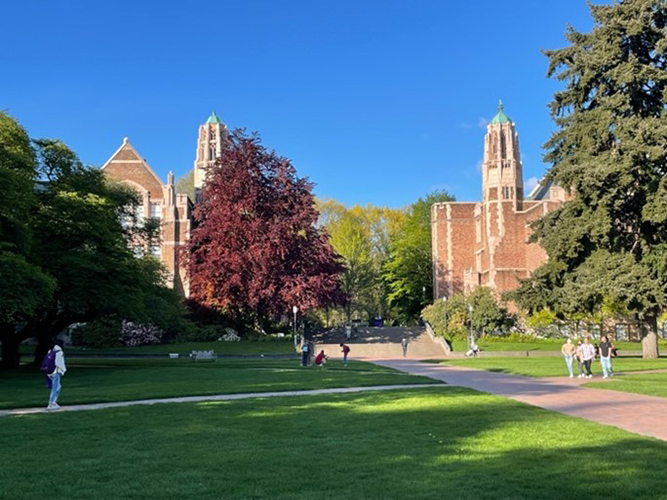
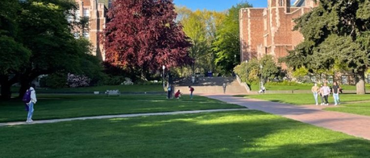
University of Washington campus.
I arrived in Seattle just in time to see the last few days of the cherry blossoms in May 2022. The city, on the US West coast, is well known for its tech innovation hub, rainy climate, and great coffee. Upon my arrival, I was warmly welcomed by Prof. Allbritton’s team at the beautiful University of Washington campus. I spent two weeks in the Allbritton lab under the mentorship of Dr. Cecilia Villegas Novoa. During the first part of my training, I learned how to generate the 3D scaffolds. This involved adapting the transwells used for the scaffolds, preparing the collagen type I crosslinking solution, and using the crypt-like protrusion stamps to create crypt shapes in collagen gels. After that, I learned how to culture human colonic epithelial cells on top of the scaffolds in order to perform experiments. Finally, I learned how to stain the cells on the scaffold and isolate whole crypts for imaging purposes.
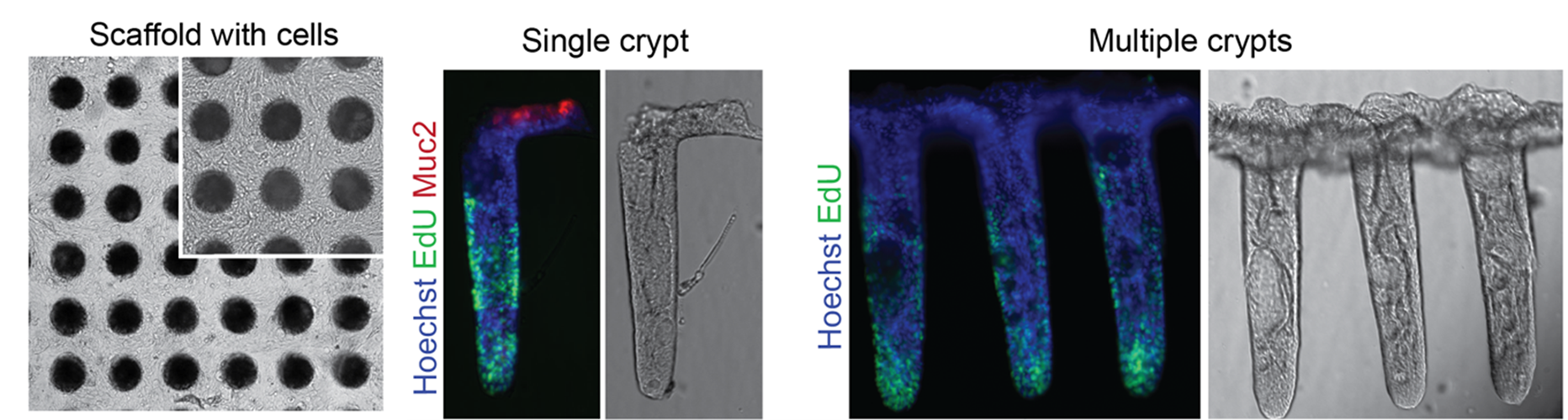
Images of the 3D crypt culture model with stained cell nuclei (blue/ Hoechst) and proliferating human colonic epithelial cells (green / Edu). Expression of Muc2 at the top of the crypt is part of the intestinal mucus barrier (Red).
A promising 3D cell model for CRC research
The advantage of this 3D crypt model is that it provides a better representation of the intestinal stem cell niche, especially by creating compartments that allow the confinement of human colonic epithelial cells. The culture system also provides opportunities to impose a gradient of signaling factors that allows for polarization of the cells, with the stem cells maintained at the bottom of the crypt. Moreover, the cell model enables us to easily manipulate factors such as crypt size and signaling factors to study their effect on intestinal stem cells.
We have now successfully implemented this 3D crypt cell model in our lab at Cancer Center Amsterdam. We aim to study several phenomena related to CRC such as the impact of oncogenic mutations on stem cell dynamics, the role of the niche signalling and niche size on stem cell maintenance, and assess the feasibility of performing high-throughput drug screens.
Acknowledgements
I would like to greatly thank Cancer Center Amsterdam for giving me the opportunity to travel to Seattle and get hands-on experience from other experts in this field. I would also like to thank Prof. Allbritton and her team for welcoming me in their lab and taking the time to share their expertise. I am grateful for all the insights and knowledge gained from this short trip.
For more information, contact Rana Ramadan.
References
- Ramadan, R., van Driel, M. S., Vermeulen, L., & van Neerven, S. M. (2022). Intestinal stem cell dynamics in homeostasis and cancer. , (5), 416–425. https://doi.org/10.1016/j.trecan.2022.01.011
- Wang, Y., Kim, R., Gunasekara, D. B., Reed, M. I., DiSalvo, M., Nguyen, D. L., Bultman, S. J., Sims, C. E., Magness, S. T., & Allbritton, N. L. (2018). Formation of Human Colonic Crypt Array by application of chemical gradients across a shaped epithelial monolayer. , (2), 113–130. https://doi.org/10.1016/j.jcmgh.2017.10.007
Text and pictures by Rana Ramadan.
This article was created for Cancer Center Amsterdam.