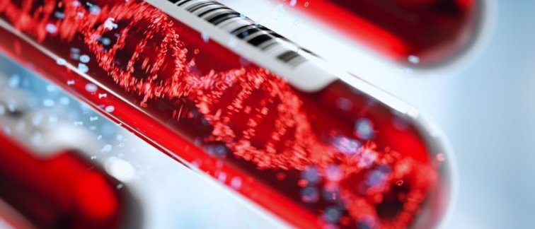
Cell-free DNA (cfDNA) circulating in the blood plasma originates from dying cells and cells that actively secrete these short fragments of nucleic acid. The average size of cfDNA was thought to be around 170 base pairs (bp) in length, but startling new evidence just published in the journal Genome Research is redrawing the landscape of cfDNA by the discovery of a new population of “ultrashort” cfDNA about 50 bp in length. The report was the result of a collaboration between Amsterdam UMC, University of Cambridge, and University of Cologne.
Hidden in plain sight
Florent Mouliere, Assistant Professor of Pathology at Cancer Center Amsterdam and senior co-author, says their work shows that the nature of cfDNA has not been completely characterized. “These ultrashort cfDNA molecules were inaccessible to conventional lab methods. Here, we show that by combining high-affinity magnetic beads for DNA isolation and single-stranded DNA sequencing methods, a previously hidden cfDNA population, sharply-organized at 50 bp, can be detected.”
Diagnostic potential
These ultrashort fragments are comprised of double-stranded DNA and ssDNA molecules, and originate mostly from hematopoietic cells. The research team wondered if the ultrashort cfDNA could have a diagnostic potential in cancer. “We found a significant difference in the abundance of ultrashort cfDNA between cancer patients and healthy individuals,” says Irena Hudecova, Research Associate at the University of Cambridge and co-first author. “The spike at 50 bp is dominant in healthy individuals, but progressively diluted in liquid biopsy samples from patients with cancer.”
Traces of tumor
The ultrashort cfDNA fragments were found to contain tumor-derived signals, but not in greater abundance when compared to other fragment size ranges. Dr. Mouliere: “By size-selecting these ultrashort fragments and analyzing copy number alterations in cancer patients, we observed that they harbor tumor DNA in equal fraction to longer cfDNA fragments bounds to nucleosomes.”
Where are they coming from?
Analysis of the nucleotide composition of the ultrashort cfDNA suggest they may originate from open chromatin regions in blood cells that could be linked to transcription start sites. “The ultrashort cfDNA fragments map to accessible chromatin regions of hematopoietic cells, particularly in promoter regions with the potential to adopt G-quadruplex DNA secondary structures,” says Dr. Hudecova.
However, the biological mechanisms that produce ultrashort cfDNA fragments are still unknown, as is what causes the relative decrease in abundance in cancer patients compared to healthy individuals. “For instance, the presence or altered activity of various plasma nucleases in cancer patients could explain the lower representation of G-quadruplex sequences in ultrashort plasma cfDNA,” says Dr. Hudecova.
“The fact that the proportion of ultrashort cfDNA differs between samples from healthy individual and cancer patients means this new population of cfDNA can further improve our ability to identify samples from individuals with cancer, but further developments are needed to fully realize its clinical potential,” concludes Dr. Mouliere.
For more information contact Dr. Florent Mouliere (f.mouliere@amsterdamumc.nl) or read the scientific publication: Hudecova, I., et al. (2021) Characteristics, origin, and potential for cancer diagnostics of ultrashort plasma cell free DNA. Genome Research. Doi: 10.1101/gr.275691.121
Link to open access publication:
https://genome.cshlp.org/content/early/2021/12/20/gr.275691.121.abstract
Involved researchers from Cancer Center Amsterdam, Amsterdam UMC: Florent Mouliere.
Lab website: https://moulierelab.org/
Text by Laura Roy

