High-end CT-scanners
The Amsterdam UMC, locations AMC and VUmc, are in the possession of multiple high-end conventional and dual-energy (dual-source, TwinBeam, kVp-switching) CT scanners from Siemens (Siemens Healthcare, Forchheim, Germany) and GE (General Electric Healthcare, Waukesha, WI). Also a total body weight-bearing cone-beam CT scanner and an extremity weight-bearing cone-beam CT scanner are present, both from Planmed Oy (Helsinki, Finland). Clinical use of different CT-techniques enables a patient-specific routing to the right CT scanner. In this way optimal CT protocols are chosen in terms of indication, radiation dose, image quality and spectral separation.
Recent CT developments improved overall image quality, speed-up acquisitions times and reduced CT radiation dose while enabling more advanced analyses.
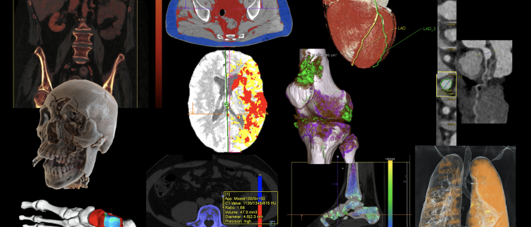
Advanced postprocessing
CT postprocessing software enables the reconstruction, evaluation and visualization of CT images, thereby improving diagnosis and clinical decision-making. Use of in-house developed software and clinically available software platforms such as syngo.via facilitate imaging workflows and reduce postprocessing time, thereby optimizing the clinical value of CT in daily practice. Commercial software applications are optimized for analysis of brain perfusion, cardiac imaging and metal artifact reduction in skeletal imaging. In-house developed applications include AI based cardiovascular diagnostic tools and dedicated software for surgical planning, joint motion analysis and knee prosthesis loosening.