1.5T&3T at Amsterdam UMC
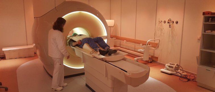
MRI is a non-invasive technique offering a unique view of living tissue and organs. MRI has applications in a wide range of (patho)physiological research areas such as the neurovascular, muscular and oncologic field, and is of high value for clinical diagnostics. Amsterdam UMC has several 1.5T and 3T Philips and Siemens MRI scanners.
Technical advances in MRI are primarily driven by the needs of medical professionals and patients. Novel MRI methods provide acceleration of scan time and advanced reconstruction of the MR data. This diminishes the burden of undergoing MRI for patients or subjects, while still providing high resolution images. Apart from standard static MRI, other techniques such as MR elastography and dynamic MRI are available. Quantitative analyses of MR data enable objective imaging of biomarkers reflecting the underlying processes in the body. These biomarker are important for clinical research, diagnostics and evaluation of treatment.
7T at Spinoza Centre
The Spinoza Centre for Neuroimaging facilitates (clinical) research using ultra-high field 7 Tesla MRI. In the brain, 7T MRI facilitates structural, functional (e.g., brain activity), connectivity, and spectroscopy imaging with increased detail compared to conventional MRI scanners. Imaging in different parts of the body (e.g., liver or calf) or different nuclei (e.g., 23NA or 31P) are also possible at 7T. For more information, see https://www.spinozacentre.nl.
Links
- Research MRI scanner software patch enabling PROspective Undersampling
in Multiple Dimensions (PROUD) on a cartesian k-space grid, see https://www.amc.nl/web/leren/research-62/research/software.htm - MRI Research Amsterdam UMC: https://mriresearch.amsterdam/