Better, individualized treatment is desperately needed for people with esophageal cancer. Current treatment usually consists of an intensive course of radiation and chemotherapy followed by surgery. Surgery comes with risks: many patients have serious side effects and require longer hospitalization. However, approximately 1 in 4 tumors has completely disappeared as a result of radiation and chemotherapy, making surgery and the risk of additional complications unnecessary.
How can we tailor treatment?
Dr. Oliver Gurney-Champion thinks better imaging is the answer. With funding from the Dutch Cancer Society (KWF Kankerbestrijding), Dr. Gurney-Champion will lead a team that will develop better magnetic resonance imaging (MRI) techniques) that can “see” how effective the radiation and chemotherapy treatments are and prevent risky surgery in people for whom the tumor has already been cured.
Notoriously poor
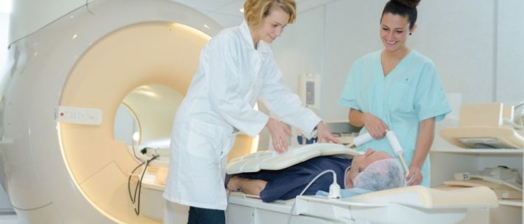
MRI uses strong magnetic fields to create detailed images of tumors and their surrounding tissues. Currently, MRI imaging of esophageal tumors is only performed in a few expert university medical centers. But sometimes, even experts cannot interpret the images due to notoriously poor image quality. This is partly due to fact that esophageal cancer is located in an area where there is a lot of movement due to breathing and heartbeat. Also, esophageal tumors are relatively small, making them difficult to accurately image with current MRI technology.
Novel MRI technologies
Dr. Gurney-Champion explains: “Together with experts in the fields of oncology, surgery, pathology, radiotherapy and radiology, I will develop novel MRI technologies that makes it possible to better visualize esophageal tumor pathophysiology that can help personalize the treatment of this cancer. Using Artificial Intelligence, I will develop motion-compensated free-breathing image acquisition with substantially improved image quality. With better imaging that can be interpreted even by non-experts, we take an important step towards selecting the optimal treatment of esophageal cancer for any patient at any hospital.”
Blood flow and density
The proposed techniques enable quantitative measurements and can visualize specific features of esophageal cancer such as blood flow and density of tumor cells. “Good blood flow is essential for chemotherapy to reach all esophageal cancer cells. By characterizing blood flow, it will be possible to predict whether these treatments will work even before radiation and chemotherapy,” said Dr. Gurney-Champion. “Tumors often have a higher cell density. So, response to therapy can be determined by measuring a decrease in cell density over time.”
Tailoring treatment through rational choices
“We expect that this new high-quality quantitative MRI could have an important clinical impact, as it enables rational choices for starting, modifying or continuing the most optimal therapy. It can also help develop new therapies by accurately evaluating therapy effectiveness at an early stage,” says Dr. Gurney-Champion. “However, before our newly developed MRI techniques can be applied in daily clinics, larger studies need to be done to confirm that the imaging can indeed predict effectiveness of the various treatment options.”
For more information, contact Oliver Gurney-Champion