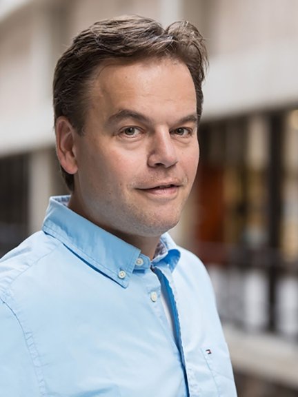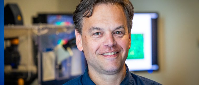
Eric Reits, Professor of Cellular Imaging, much rather describes himself as a man with two great ‘hobbies’: Huntington’s disease (HD) and microscopy. It is clear that he has a deep passion for these two specialisms, demonstrated by the substantial funding he recently received as coordinator of both the CureQ and the NL-BioImaging AM (NL-BI) consortium.
Cellular Imaging core facility
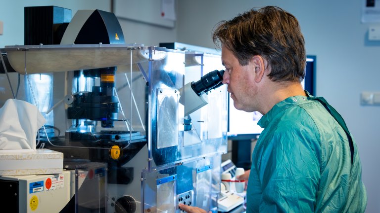
You will usually find the professor at the Cellular Imaging core facility, either in his office or immersed behind his favorite light microscope: a confocal laser-scanning microscope. To reach the core facility, simply use the elevator located between building part L and M, and head to the third floor – this kind of feels like getting transported to Willy Wonka's magical world. But instead of chocolate rivers, you are encountering a world of cutting-edge microscopy with a wide range of instrumentation for electron and fluorescence microscopy and flow cytometry. The electron microscopes enable high resolution 2D and 3D images of fixed neurons, organoids and brain tissues, while especially the confocal microscopes enable visualization of processes in living neurons, brain organoids and small animal models such as zebrafish.
All these resources are accessible to researchers at Amsterdam UMC. Simply request access via the website and schedule your session. To get you all set up, the core facility provides individual hands-on training on instruments and methodologies, data-analysis, and discussion of data, but also courses on basic and advanced microscopy, data-analysis, and converting microscopy images to publication-grade figures.
Revolutionizing microscopy
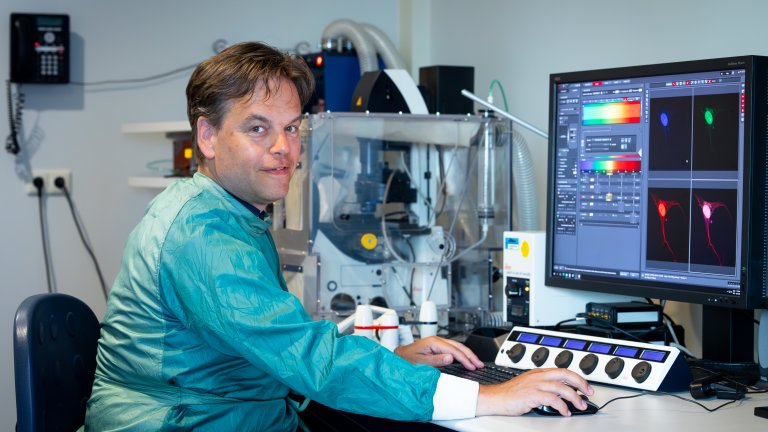
Currently, Reits is at the forefront of expanding the core facility by developing an advanced state-of-the-art light microscope together with all 18 Dutch universities, university medical centers, and thematic research institutes including the NKI and Hubrecht. Reits' preferred microscopes provide either rapid ‘functional imaging’ of processes such as calcium signaling in single cells using fluorescent biosensors, or very slow testing thousands of therapeutic compounds in large multiwall plates with neuronal cells. “When evaluating 10,000 potential medications on Huntington patient-derived neuronal cells using fluorescent biosensors, I need a microscope that can simultaneously and rapidly measure how neurons are communicating with each other in 400-500 different groups of cells, and whether particular compounds or treatments make them to behave like healthy neurons again.” The proposed innovative microscope eliminates the compromise between speed and quantity.
Additionally, they aim to integrate Artificial Intelligence and machine learning, so the microscope automatically analyzes and interprets images, recognizes patterns, and makes informed decisions based on the acquired data during the imaging (called on-the-fly analysis). “During measurement, you want the microscope to recognize what you want to observe and make decisions such as isolating cells of interest for subsequent analysis by genomics or proteomics,” the professor explains. While it may take up to two years, this globally unique self-steering or ‘smart’ microscope, will find its home at the core facility Cellular Imaging. Different microscopy companies and expertise groups within NL-BI are involved in the development of the proposed microscope.
Bridging the gap
As a true connector, Reits is capable of uniting large, varying groups of people. In his pursuit for a therapy for Huntington's disease, he brings together Dutch medical centers, biotech companies, universities of applied sciences and patient foundations. This consortium (www.cureQ.nl) will generate HD patient-derived neuronal iPSC models to perform numerous measurements using a wide variety of imaging and omics techniques, ultimately resulting in a diverse array of datasets and AI-derived predicative models that will enable academic and industry researchers to test new therapeutic strategies, and neurologists to predict age-of-onset more accurately.
Simultaneously, this presents us with an entirely new puzzle to unravel. The professor explains: “I have seen large projects fail because they are not able to merge datasets from different medical centers, or from different technologies or core facilities. Eventually, we will all face the same problems, and these can only be resolved if we work together.” Eric's vision underscores that this intricate landscape can’t be navigated alone; it requires the fusion of people, techniques, and exchanges to achieve their collective goals.
The importance to store and connect microscopy and omics data
Images of communicating neurons are generated by the microscope facility, the proteomics facility reveals the production and modifications of proteins, the metabolomics facility determines alterations in homeostasis and metabolism, and the genomics facility deciphers RNAs – in short, all these Amsterdam UMC core facilities are creating different kinds of data output. “To be able to link this data for subsequent AI analysis, we need an approach as straightforward as organizing an Excel sheet,” emphasizing the need for simplicity in data unification. “That is why we are assembling a core-facility embedded research team with a lot of talented young scientists of universities of applied sciences, who are specialized in AI, data analysis and management,” Reits says. After connecting the different datasets, Artificial Intelligence programs can carefully analyze it, uncovering hidden patterns that determine disease onset and progression that were not visible before. Only imagine the possibilities that this groundbreaking method could unlock, not only for conditions like Huntington's disease and other neurological disorders, but also for the advancement of cancer research, cardiovascular diseases, rare genetic conditions, and numerous other areas in the medical sector.
Pursuing future solutions
Essentially, the pioneering professor is actively pursuing solutions to future challenges. Drawing from his passion for microscopy and his deep interest in Huntington's disease, his primary goal is to help carriers of the Huntington’s disease gene. As co-founder of Campagneteam Huntington (www.doodgezwegen.nl) he meets frequently with members of affected families. “If all goes as planned, we will be able to accurately predict the age-of-onset for each gene-carrier and start in time with therapeutic treatments to delay or prevent onset of HD,” Reits clarifies. Here is where it all began, as the communication between two Amsterdam UMC facilities, metabolomics and microscopy, plays a crucial role in achieving this goal. A challenge that has transformed into Eric Reits' significant ideas and actions. Throughout his journey, one certainty remains: Eric Reits is wholeheartedly devoted to finding a cure.
In short
What is it?
A SMART microscope, unique on a global scale.
What can I do with it?
Rapidly study numerous neuronal organoids combined with on-the-fly analysis to recognize specific phenotypes, and data-driven decision-making by the microscope.
Who can use this?
All researchers within Amsterdam UMC, but you will most likely run into PhD Candidates.
Where is it?
At Amsterdam UMC, location AMC, on L3: the third floor of building part L.
What is the ideal spa day for the new microscope and the core facility in general?
Providing effective trainings to users, state-of-the-art instruments and continually optimize software for self-steering microscopy, data management and analysis.
How can I utilize the technology at Cellular Imaging?
Go to https://www.cellularimaging.nl/agenda/ and follow the instructions.
Who is the contact person?