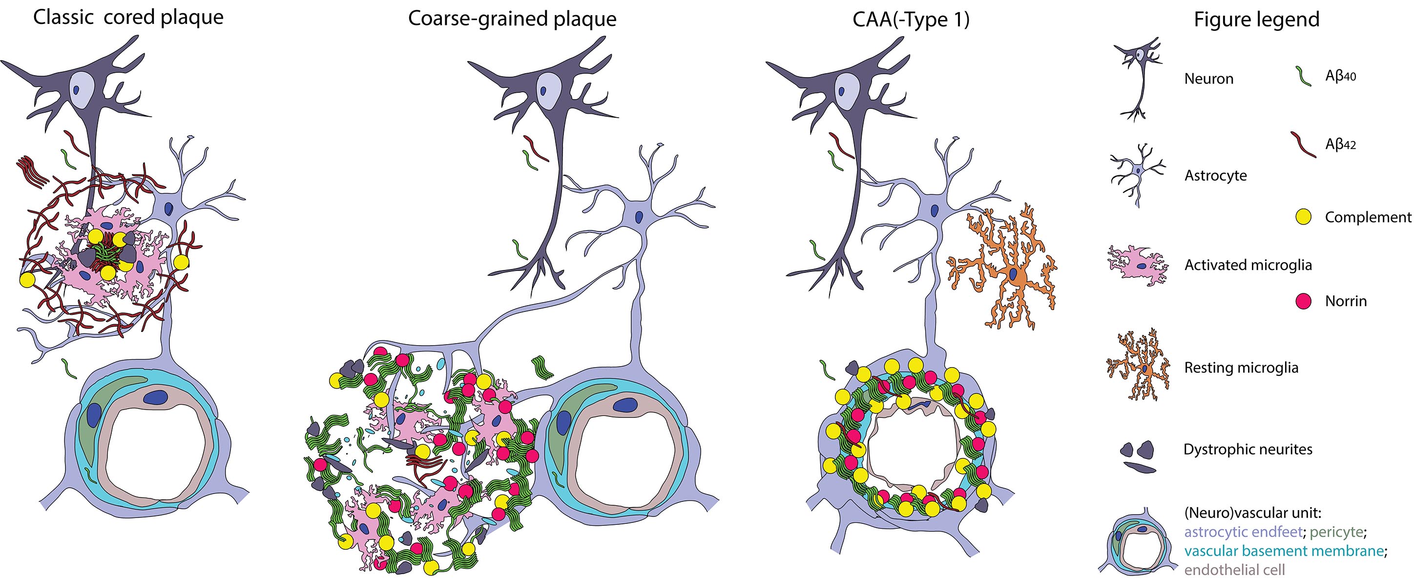
This coarse-grained plaque is observed more frequently in Alzheimer’s disease cases with an early disease onset. It contains different amyloid proteins typically observed in plaques, and is associated with vascular changes and a strong neuroinflammatory response. The results support that patients with these plaques need different amyloid-targeted therapies.
Coarse-grained plaque in a cohort study
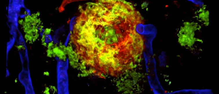
Baayla Boon together with Marjolein Bulk characterized and semi-quantitatively scored the coarse-grained plaque in a cohort of 74 post-mortem brain specimens. Their cohort was comprised of early-onset AD, late-onset AD, and cases without dementia but positive for amyloid deposits in the brain. The coarse-grained plaque is with a diameter between 30-100 μm larger than the classic cored plaque. At first glance the coarse grained plaque looks similar to the cotton wool plaque, a characteristic deposit seen in cases with specific PSEN1 mutations. However, with their multiple cores, pores that are oddly devoid of Aβ, tube-like or trabecular structures running through them, and a vague border, the coarse-grained plaque really has a different morphology. They stain for APP and prion protein, which could indicate damage to adjacent neurites. Coarse-grained plaques contain mostly Aβ40, though in the larger ones, this Aβ40 seemed to surround an Aβ42 core, which is quite the opposite for classic cored plaques. Glial cells infiltrate the coarse grained plaque in a pattern which is distinct from other plaque varieties.
Direct connection
Whereas 66% of late-onset and even 95% of early-onset cases were positive for coarse-grained plaques, the plaque was absent in cognitively normal cases. This means the plaque is associated with the onset of clinical symptoms for Alzheimer’s disease. Interestingly, the coarse-grained plaque was associated with the genetic risk factor APOE ε4 and was always observed in cases homozygous for the ε4 allele. Using 3D confocal laser scanning microscopy, the authors showed that 84% of the coarse-grained plaques was in direct contact with a bloodvessel. Besides their direct connection with the vessels, the plaques also contained laminin, a component of blood vessel walls and norrin, a protein recently discovered by the same group to be associated with the vascular-type of Alzheimer’s disease, also referred to as CAA. The authors hypothesize that these coarse-grained plaques mostly comprised of Aβ40 aggregate at the border of the blood-brain barrier and the brain parenchyma (see the figure below). Due to their difference in composition and location within the brain parenchyma, these plaques might need a different therapeutic approach than the common dense-cored plaques or the cerebral angiopathy associated amyloid.
This study was the result of the close collaboration between different research groups within Amsterdam Neuroscience, the Netherlands Brain Bank, Leiden University Medical Center, University of Bonn and the University of Rhode Island, and is part of the by ZonMw Memorabel funded project ‘PAGE-AD’. The results are published in Acta Neuropathologica, read the article of Boon et al.