Pioneers
Along with physician and researcher Yousif Dawood, De Bakker succeeded in imaging the embryo in great detail with a new technique called micro-CT. This imaging technique is applied in geology to examine rocks, and food technologists also use micro-CT to assess the texture of food. De Bakker stated in the newspaper: “We are among the pioneers who utilize micro-CT to study human tissue.”
On regular CT scans, only the relatively large structures in tissues are visible, says De Bakker. “But micro-CT turns out to be very suitable for soft tissue imaging if the contrast dye has plenty of time to spread everywhere. We can now scan details as small as a micrometer. I predict that in ten years all hospitals will have this kind of device for pathological examination. After all, this is a huge improvement over the usual time-consuming microscopic examination.”
Understanding how a miscarriage occurs
De Bakker and Dawood will make more such scans of embryonic tissue in the coming year. “This can yield a wealth of information for us about the development of early embryos up to approximately 1 cm in length, as well as the yolk sac, the amniotic membranes, and the placenta. We hope to use this knowledge to gain a better understanding of how an ectopic pregnancy or early miscarriage occurs.”
The response to the images of the embryo and the technology behind it has been overwhelming. Requests for cooperation and information are coming from all over – from specialists in virtual reality, for instance, as well as from other doctors or researchers.
A cooperative study has already begun. De Bakker and Dawood will team up with scientists at the University of Leuven to develop new techniques for research on the origins of organ defects, among other things. Additionally, they will be focusing on new ways to conduct research on deceased fetuses. Many parents do not want their deceased child to undergo a post-mortem examination. An imaging technique such as a scan may be an option to nevertheless obtain a considerable amount of information on the possible defect or cause of death.
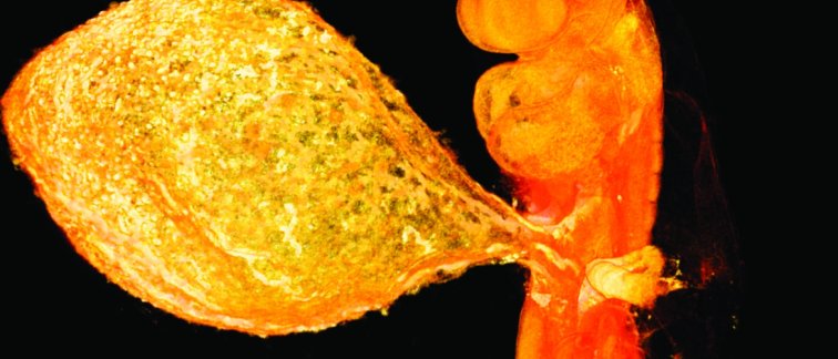
Award for most striking image
In the picture and video, you can see the embryo with a yolk sac attached. The tissue surrounding it is from the placenta. The publication about it in the journal Radiology received the award for the best image of the year. “We were completely taken by surprise. Radiology is the largest and most well-known journal in the field. The fact that we were awarded the grand prize is truly exceptional.”
3D atlas
De Bakker previously imaged the developing human embryo in a three-dimensional atlas. For this purpose, images of more than 15,000 sections of embryos between the ages of 15 days and 2 months were digitalized. The embryos are from the historic Carnegie Collection in the United States. The atlas had already cleared up several misconceptions about the development of the human embryo, leading to a publication in the renowned scientific journal Science.
Source: NRC
Images: Dawood Y, de Bakker BS., Amsterdam UMC, Radiology. 2020 Oct;297(1):32.