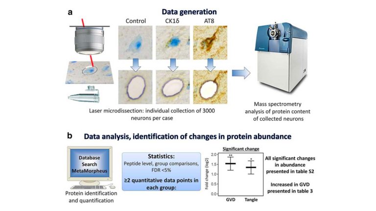
Granulovacuolar degeneration (GVD) is a common feature in Alzheimer’s disease (AD). The occurrence of GVD is closely associated with that of neurofibrillary tangles (NFTs) and GVD is even considered to be a pre-NFT stage in the disease process of AD. However, the composition of GVD bodies, the mechanisms associated with GVD and how GVD relates to NFTs is not well understood. By combining immunohistochemistry (IHC) and laser microdissection (LMD) the authors isolated 3000 neurons each with GVD or tangles separately from human post-mortem AD hippocampus.
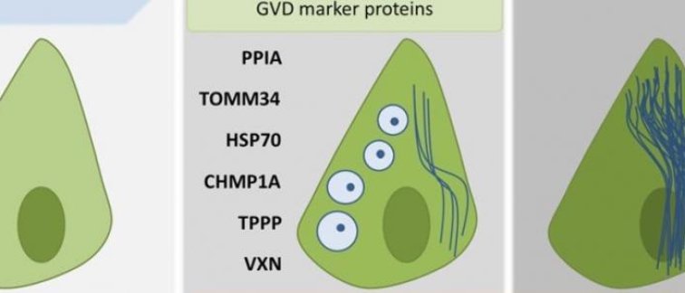
Due to the high resolution of the analysis of specific neuronal populations the study identified many proteins that had not been associated previously with GVD or NFTs. The data show that GVD and neurofibrillary tangle bearing neurons are molecularly different but closely related. GVD bearing neurons have increased presence of proteins associated with protein folding, endolysosomal function and glycolysis, while there is a decrease of proteins involved in RNA processing and proteasome components. Increased levels of proteins in GVD that promote protein folding, synaptic plasticity and neurogenesis are in line with previous functional data (Wiersma et al. Acta Neuropath. 2019, Scheper Lab, CNCR), showing that tau pathology induces GVD. The data support the model that GVD is part of a pre-NFT stage representing a phase in which proteostasis and cellular homeostasis is disrupted.
Read the article published in Acta Neuropathologica ‘The proteome of granulovacuolar degeneration and neurofibrillary tangles in Alzheimer’s disease’.