Vera Keil
Neuro-oncological imaging, the imaging of brain tumors, is a diversified field of research at Amsterdam UMC where several smaller labs work constantly together like the fingers of a hand to get a grip on the disease. My own team is closely linked to the teams of professor Frederik Barkhof and Dr. Henk Jan Mutsaerts, and currently follows two main research questions:
1. How can we reduce the necessity to inject gadolinium-based contrast agent for brain tumor imaging? and 2. What role can advanced MRI techniques play in redefining imaging-based diagnostics and therapy planning?
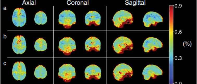
Advanced MRI is a term that describes all kinds of MRI sequences that are not part of the standard repertoire of T1- and T2-weighted images and their variation. Often these images deliver quantifiable data such as on chemical properties of a tumor or on its perfusion. As part of our research, we focus on all currently established perfusion techniques (DSC, DCE, ASL) as well as experimental variations (IVIM, blood-brain barrier sensitive ASL) and CEST imaging.
Our spearpoint studies are GLIOCARE (funded by the Hanarth Fonds) and REDUCE (funded as an Innovation project of AUMC). We could recently show that the availability of post-gadolinium-contrast sequences does not improve the radiologists’ diagnostic accuracy in predicting glioma subtypes, and that non-contrast sequences can predict the contrast enhancement quality and quantity with a high reliability. Our next goal is to study the impact of a 50% dose reduction on perfusion image results and standard image quality to maybe soon be able to reduce the gadolinium contrast dose for all brain tumor patients. This project has a high environmental impact.
Our group further applies deep learning and radiomics methods to investigate the information content of advanced MRI methods for applications such as alternative tumor resection borders and synthetic post-contrast images. As AI-based research is strongly data-driven, our researchers are currently taking the effort to launch IMAGO, Amsterdam UMC’s first glioma imaging database for public research use.
We work in a very close collaboration with other CCA groups (among these: Prof. Philip De Witt Hamer, Dr. Linda Douw, Prof. David Noske, Dr. Elsmarieke van de Giessen), but also expand our network to numerous European partners including at NKI-AvL, Erasmus MC Rotterdam, Helmholtz Center Dresden-Rossendorf, University Hospital Prague, University Hospital Ulm and University College London. Our group welcomed numerous students for short term scientific missions in the past, and keeps up a strong training and development dedication for young clinical researchers with a high proportion of female staff.
Beyond brain tumor imaging, I personally also focus on quantitative imaging for other conditions, such as Multiple sclerosis in the REDUCE and TACTIX projects, and advanced relaxometry in a collaboration with UMC Utrecht, group Sbrizzi.
Latest news:
- Our team won the EUCAIM Open Call and will publish the Amsterdam Glioma Database IMAGO this Summer!
- together with Cerebriu S/A (link: Cerebriu | Expand MRI accessibility through advanced AI) we won a TKI grant (SECURO) and will develop a tool to reduce contrast agent in brain MRI.
Group members
PhD students:
- Ivar J. Wamelink
- Aynur Azizova
- Rajeev A. Essed
- Joao Ramos
- Alex Ferles
- Kati Farkas
- Selena Huisman
Current research students:
- Nathalie Ringrose (voluntary)
- Dr. Afolabi Ogunleye (CAMERA network)
- Olga Filipowicz (research stage, BA)
Former members:
- Sharon Vollenga (MA Leiden)
- Dr. Shuncong Wang (freelance researcher)
- Marcus Cakmak (honors student)
- Elif Kaya (Erasmus+ program, 2023)
- Basak Usta (Erasmus+ program, 2024)
- Hazar Tuze (Erasmus+ program, 2024)
- Laura Cristobal Saez (Erasmus stagiaire 2024)
- Norman Kornemann (Bracco fellowship)
- Gulce Turhan (COST Action mobility grant)
- Ayse Irem Cetin (COST Action mobility grant)
- PD. Dr. Nico Sollmann (COST Action mobility grant)
- Maarten Balder (voluntary)
- Elske Korebrits (BA)
PhD publications
Key publications
Wamelink IJHG, Azizova A, Booth TC, Mutsaerts HJMM, Ogunleye A, Mankad K, Petr J, Barkhof F, Keil VC. Brain Tumor Imaging without Gadolinium-based Contrast Agents: Feasible or Fantasy? Radiology. 2024 Feb;310(2):e230793. doi: 10.1148/radiol.230793. PMID: 38319162; PMCID: PMC10902600.
Wamelink IJHG, Kuijer JPA, Padrela BE, Zhang Y, Barkhof F, Mutsaerts HJMM, Petr J, van de Giessen E, Keil VC. Reproducibility of 3 T APT-CEST in Healthy Volunteers and Patients With Brain Glioma. J Magn Reson Imaging. 2023 Jan;57(1):206-215. doi: 10.1002/jmri.28239. Epub 2022 May 28. PMID: 35633282; PMCID: PMC10084114.
Wamelink IJHG, Hempel HL, van de Giessen E, Vries MHM, De Witt Hamer P, Barkhof F, Keil VC. The patients' experience of neuroimaging of primary brain tumors: a cross-sectional survey study. J Neurooncol. 2023 Apr;162(2):307-315. doi: 10.1007/s11060-023-04290-x. Epub 2023 Mar 28. PMID: 36977844; PMCID: PMC10167184.
Azizova A, Prysiazhniuk Y, Wamelink IJHG, Petr J, Barkhof F, Keil VC. Ten Years of VASARI Glioma Features: Systematic Review and Meta-Analysis of Their Impact and Performance. AJNR Am J Neuroradiol. 2024 Aug 9;45(8):1053-1062. doi: 10.3174/ajnr.A8274. PMID: 38937115; PMCID: PMC11383402.
Essed RA, Prysiazhniuk Y, Wamelink IJ, Azizova A, Keil VC. Performance of amide proton transfer imaging to differentiate true progression from therapy-related changes in gliomas and metastases. Eur Radiol. 2024 Aug 12. doi: 10.1007/s00330-024-11004-y. Epub ahead of print. PMID: 39134744.
Contact
Keywords
Advanced MRI | glioma | brain tumor | gadolinium | quantitative imaging